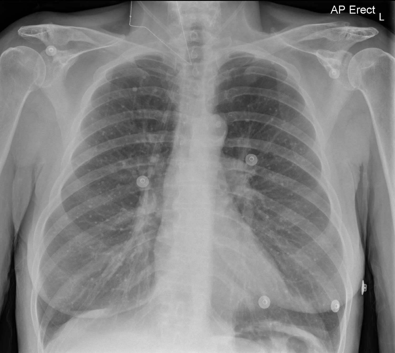
 Calcified granuloma definition, causes, symptoms, diagnosis & treatment
What you need to know about calcified granulomas A calcified granuloma is a specific type of tissue inflammation that has been calcified over time. When something is called "qualified", it means it contains deposits of the calcium element. Calcium has a tendency to collect in tissue that is healing. The formation of granulomas is often caused by an infection. During an infection, immune cells surround and isolate foreign material, such as bacteria. Granulomas can also be caused by other inmunitary systems or inflammatory conditions. They are most commonly found in the lungs. But they can also be found in other organs of the body, such as the liver or spleen. Not all granulomas are calcified. Granulomas are made up of a spherical group of cells that surround the inflated tissue. Over time they can be calcified. A calcified granuloma has a bone-like density and will appear brighter than the surrounding tissue in a .As uncalcified granulomas do not contain calcium deposits, they may appear as a less different group of cells in an x-ray or . Because of this, they are often misdiagnosed as cancerous growths when seen in this way. If you have a calcified granuloma, you may not even know it or experience symptoms. Typically, a granuloma will only cause symptoms if an organ's ability to function properly is affected due to its size or location. If you have a calcified granuloma and are experiencing symptoms, it may be due to an ongoing underlying condition that caused the shape of granuloma. The formation of calcified granulomas in the lungs is usually due to infections. These can be from a bacterial infection, like . Calcified granulomas may also form fungal infections such as or . Non-infectious causes of lung granulomas include conditions such as and . Calcified granulomas may also form organs other than the lungs, such as the liver or spleen. The most common infectious causes of liver granulomas are bacterial infection with TB and . In addition, sarcoidosis is the most common non-infectious cause of liver granulomas. Certain medicines can also cause the formation of liver granulomas. Calcified granulomas can be formed in the spleen due to TB bacterial infection or histoplasmosis of fungal infection. Sarcoidosis is a non-infectious cause of granulomas in the spleen. People who have calcified granulomas may not even know they are there. They are often discovered when subjected to an image procedure such as an X-ray or a CT scan. If your doctor discovers a calcification area, you can use image technology to evaluate the size and pattern of calcification to determine if it is a granuloma. Calcified granulomas are almost always benign. However, less commonly, they may be surrounded by a cancer tumor. Your doctor may also perform additional tests to determine what caused the formation of granulomas. For example, if calcified granulomas are discovered in the liver, your doctor may ask about your medical and travel history. They can also perform laboratory tests to evaluate their liver function. If necessary, a biopsy can also be taken to confirm an underlying condition that has caused granuloma formation. Since calcified granulomas are almost always benign, they usually do not require treatment. However, if you have an active infection or condition that is causing granuloma formation, your doctor will work to treat that. If you have an active bacterial or fungal infection, your doctor will prescribe an appropriate antibiotic or antifungal. Antiparasitic praziquantel can be used to treat parasitic infection due to schistosomiasis. Non-infectious causes of granulomas such as sarcoidosis are treated with corticosteroids or other immunosuppressant medications to control inflammation. Sometimes the formation of granuloma can cause complications. Complications of granuloma formation are often due to the underlying condition that caused them. The granuloma formation process can sometimes be disruptive to the tissue function. For example, schistosomiasis of parasitic infection can cause granulomas to form around the eggs of parasite in the liver. The granuloma formation process can in turn lead to . This is when excess connective tissue accumulates in the scar tissue of the liver. This can interrupt the structure and function of the liver. If you have an active infection or other condition that leads to the formation of granuloma, it is very important to be treated to prevent any complication. If you have one or more calcified granulomas, you probably don't know you have them. If a calcified granuloma is diagnosed, granuloma may not require treatment. If you have an underlying condition or an infection that leads to granuloma formation, your doctor will work to treat that. The individual perspective depends on the condition in question. Your doctor will work with you to establish a treatment plan and address any concerns. Last medical review on February 13, 2018Read this following
GoogleI'm sorry... Google Sorry... but your computer or network can send automated queries. To protect our users, we cannot process your request right now.

A radiograph taken for the right shoulder shows old healed... | Download Scientific Diagram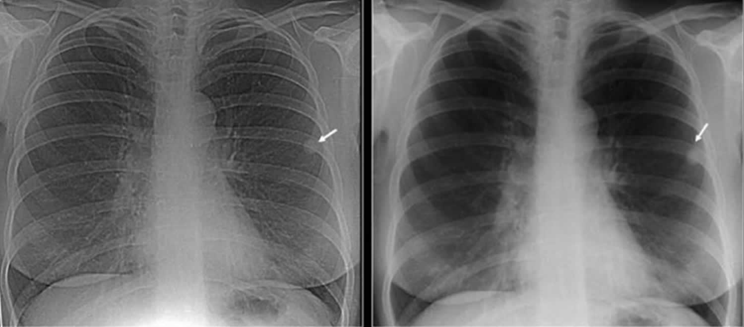
Calcified granuloma definition, causes, symptoms, diagnosis & treatment
Pulmonary Pathology
Calcified Granuloma On Chest X-Ray – Radiology In Plain English
A rare case of pulmonary hyalinizing granuloma with calcification in a 5 year old boy - ScienceDirect
How is calcified granuloma treated? - Quora
Topical Review: Pulmonary calcifications: a review
Calcified pulmonary nodules | Radiology Reference Article | Radiopaedia.org
MedPix Case - Granuloma
Calcified Granuloma: In Lung, Treatment, More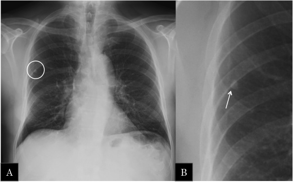
EPOS™ - C-2005
Topical Review: Pulmonary calcifications: a review
Calcified Granuloma Lung Simulates An Arteriovenous Malformation (AVM) - Chest Case Studies - CTisus CT Scanning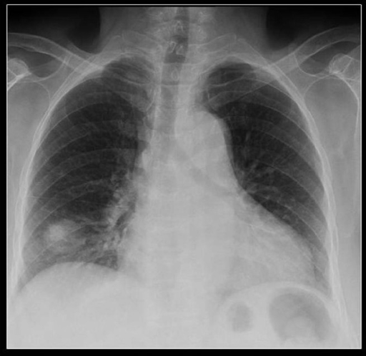
A chest radiograph showing a large PN at the right lung | Open-i
Is calcified granuloma inside the lungs dangerous? - Quora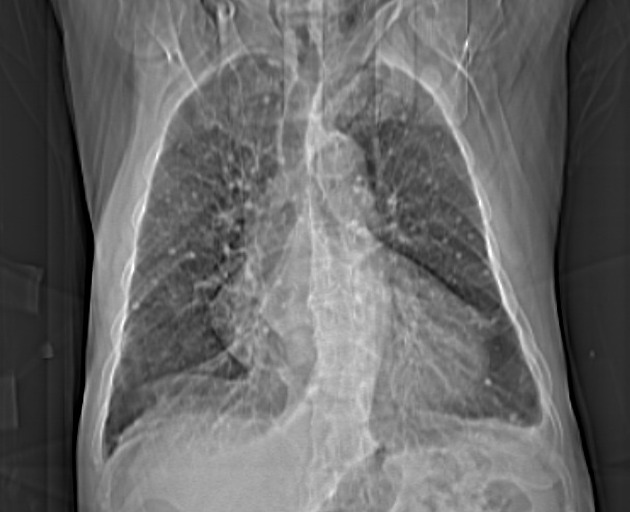
Multiple calcified pulmonary nodules | Radiology Case | Radiopaedia.org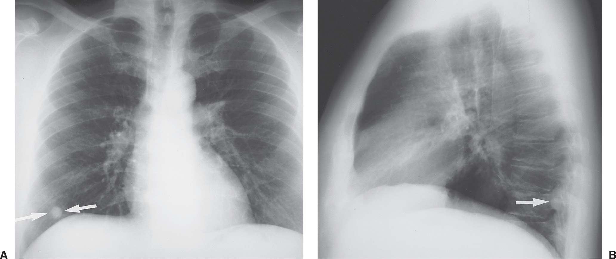
Solitary and Multiple Pulmonary Nodules | Radiology Key
Pulmonary Coccidioidomycosis: Pictorial Review of Chest Radiographic and CT Findings | RadioGraphics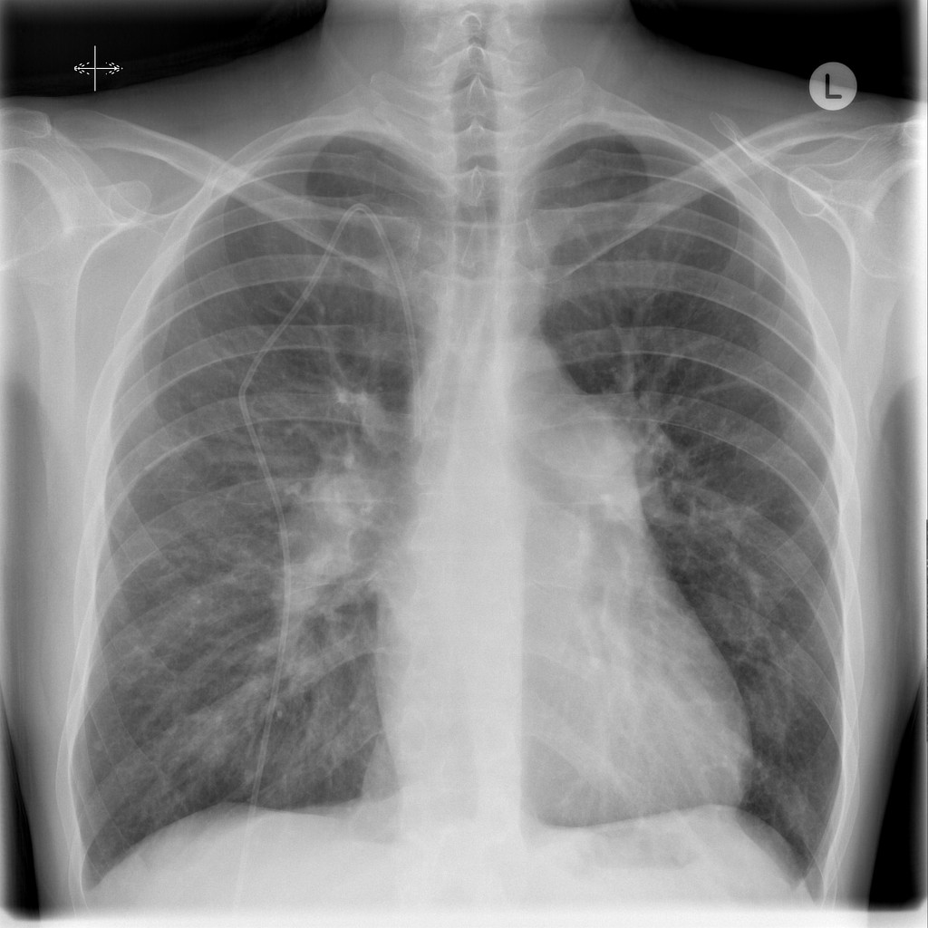
Sarcoidosis associated pulmonary arterial hypertension | Radiology Case | Radiopaedia.org
High attenuation in the lungs on CT: Beyond calcified granulomas | Lunges, High, Ct scan
Abnormalities suggestive of latent tuberculosis infection on chest radiography; how specific are they? - ScienceDirect
Differential diagnosis of granulomatous lung disease: clues and pitfalls | European Respiratory Society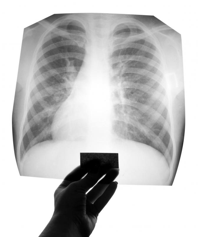
What is a Calcified Granuloma? (with pictures)
Solitary pulmonary nodule: A diagnostic algorithm in the light of current imaging technique Khan AN, Al-Jahdali HH, Irion KL, Arabi M, Koteyar SS - Avicenna J Med
Lung Granuloma|Causes|Symptoms|Treatment|Diagnosis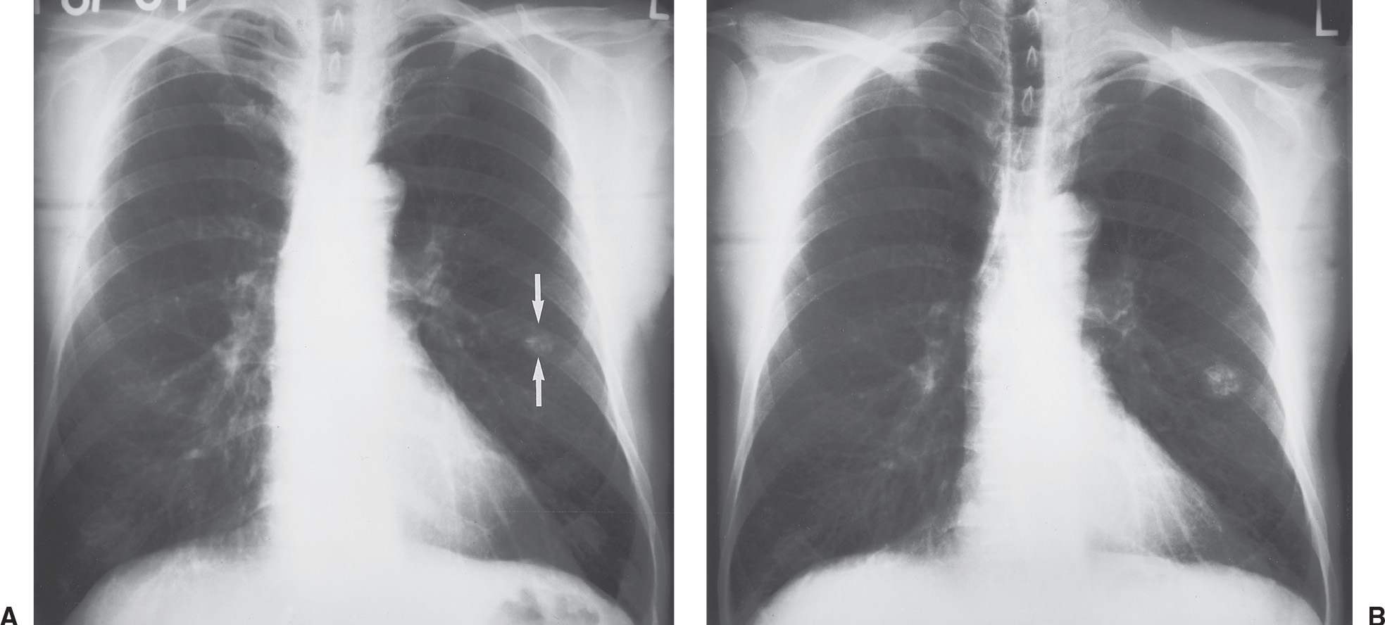
Solitary and Multiple Pulmonary Nodules | Radiology Key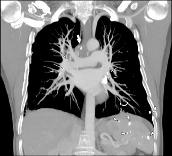
A Reminder From the Past: Unsuspected Splenic Calcified Granulomatosis Decades After Pulmonary Tuberculosis | The American Journal of Medicine Blog
Internet Scientific Publications
A chest radiograph showing calcified metastases from an osteogenic... | Download Scientific Diagram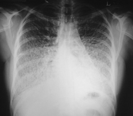
Calcified pulmonary nodules | Radiology Reference Article | Radiopaedia.org
PDF) The calcified lung nodule: What does it mean?
Calcifying fibrous pseudotumour of the lung | Thorax
The calcified lung nodule: What does it mean? - Abstract - Europe PMC
Pulmonary hyalinising granuloma: A rare cause of multiple lung nodules in lung cancer clinic - ScienceDirect
Learning Radiology - Ghon Complex, Ranke, lesion
MedPix Case - Granuloma
Tuberculosis radiology - Wikipedia
Differential diagnosis of granulomatous lung disease: clues and pitfalls | European Respiratory Society
Analysis of chest computed tomography manifestations of non-Mycobacterium tuberculosis induced granulomatous lung diseases - ScienceDirect
Management of incidental lung nodules <8 mm in diameter - Sánchez - Journal of Thoracic Disease
 Calcified granuloma definition, causes, symptoms, diagnosis & treatment


Posting Komentar untuk "what is a calcified granuloma in the lung"